
Muscular System
The muscular system is composed of specialized cells called muscle fibres. Their predominant function is contractibility. Muscles attached to bones or internal organs and blood vessels are responsible for movement. Nearly all movement in the body is the result of muscle contraction. Exceptions to this are the action of cilia, the flagellum on sperm cells, and the amoeboid movement of some white blood cells.
Muscles allow a person to move, speak, and chew. They control heartbeat, breathing, and digestion. Other seemingly unrelated functions, including temperature regulation and vision, also rely on the muscular system. The three types of muscles are Skeletal muscle, Cardiac muscle and Smooth muscle. In this article, we find out more about the muscular system and how it controls the body.
What are Muscles?
A Muscle is a soft tissue that enables contraction and tension in an animal’s body. The types of muscle are skeletal, cardiac, and smooth. Muscles in the human body are made up of muscle fibers, connective tissue, blood vessels, and nerves. Muscle tissue consists of specialized cells called muscle fibers, which contract to move the body.
Each muscle type has its own cellular components, physiology, functions, and pathology. Structurally, muscles consist of contractile fibers organized into bundles. The muscle functions include generating force for movement, maintaining posture, and supporting vital physiological processes.
The human body has over 600 muscles that makeup about 40–50% of the body’s weight. Muscles consist of parallel bundles of muscle fibers, each containing myofibrils with sarcomeres responsible for contraction. There are three main types of muscles: skeletal muscles are attached to bones and allow voluntary movements; smooth muscles which are present in internal organs control involuntary processes; and cardiac muscles which form the heart ensure rhythmic contractions for circulation. Muscles have special properties like excitability, contractility, extensibility, and elasticity.
Types of Muscles
Based on location in the human body, there are three types of muscles.
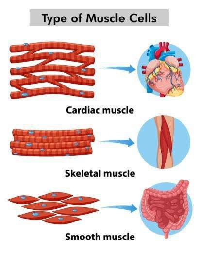
- Striated or Skeletal Muscles
- Non-Striated or Smooth Muscles
- Cardiac Muscles
1. Skeletal Muscle:
Skeletal Muscles are also known as the striated muscles and are controlled voluntarily. Skeleton muscle functions to facilitate voluntary movements and stability in the body. The skeletal muscles are mostly attached to the bones. The skeletal muscle structure consists of bundles of muscle fibers surrounded by connective tissue. The flexible muscle fibers that compose skeletal muscles can have a diameter that can vary from less than half an inch to slightly over three inches.
When these fibers contract, the muscles can move the bones, allowing humans to perform many kinds of movements. In this article, we will study the skeletal muscles structure, function, types, examples, and properties.
What are Skeletal Muscles?
The muscles are mostly attached to bones and are responsible for the body’s voluntary actions, such as walking, lifting, and running, are referred to as skeletal muscles or voluntary muscles. The skeleton muscles are multinucleated. They have a striated appearance under a microscope because of the alignment of contractile proteins. They are made up of elongated muscle fibers arranged into bundles.
Skeletal muscles work in coordination with the nervous system to perform precise and coordinated movements, providing stability and support to the skeletal structure. They are also necessary for maintaining posture and producing heat through metabolic processes.
Skeletal Muscles Definition:
Skeletal muscles are a particular type of voluntary muscle tissue that are connected by tendons to the bones and are responsible for posture maintenance and movement of the body. The arrangement of actin and myosin filaments within their fibers gives them a striated appearance.
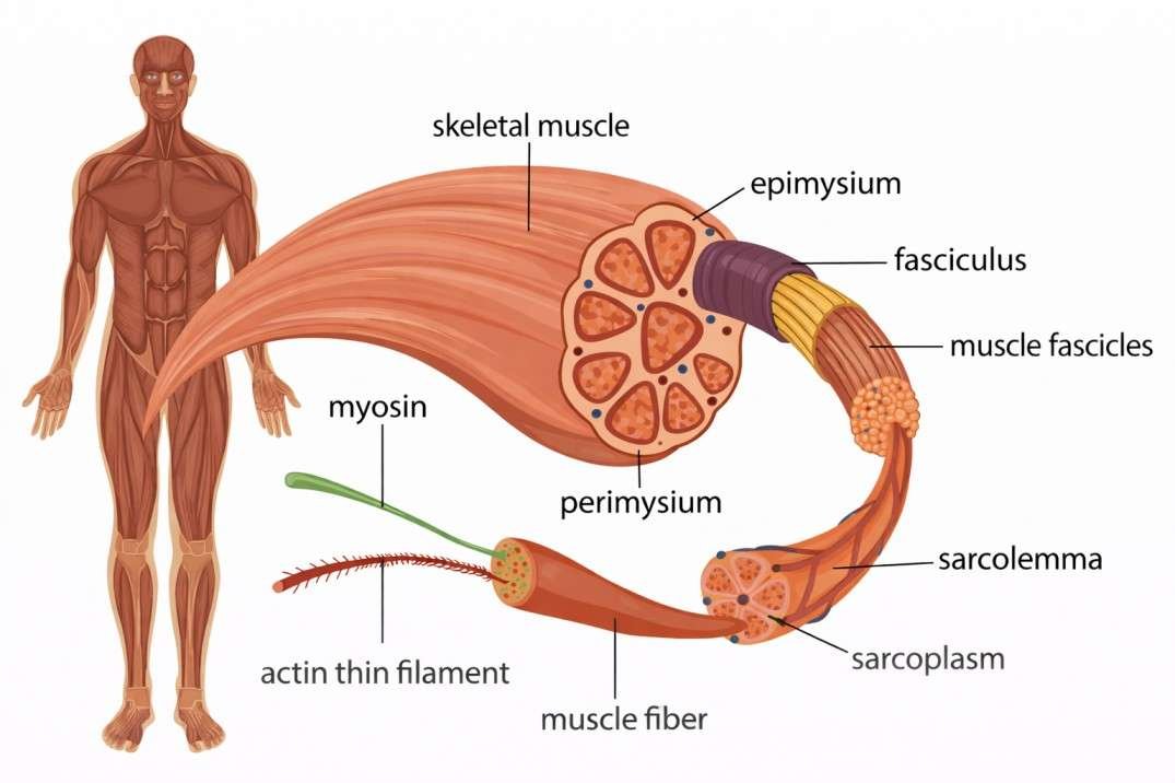
Skeletal Muscles Diagram:
The following is a labeled diagram of skeletal muscles:
Skeletal Muscles Structure:
Skeletal muscle histology examines the microscopic structure and organization of skeletal muscle tissue, highlighting its cellular composition and arrangement. Muscle fibers, the basic building blocks of skeletal muscles, are arranged in a structured manner. Long, multinucleated cells known as muscle fibers include myofibrils, which are made up of repeating units known as sarcomeres.
Skeletal muscle tissue has a uniquely striated appearance due to these sarcomeres, which are made up of overlapping actin and myosin filaments. The perimysium, a type of connective tissue, envelops the bundles of muscle fibers known as fascicles.
A full muscle is formed by the enclosure of these fascicles in an epimysium-like sheath of connective tissue. Collagen fibers from the endomysium, perimysium, and epimysium combine at the ends of muscles to produce tendons, which attach muscles to bones. The structural foundation required for force production and transmission during muscle contraction is provided by this hierarchical arrangement, allowing the body to function properly and move around.
Skeletal Muscles Functions:
The below are the functions of skeletal muscles:
- Movement: Skeletal muscles enable voluntary movements such as walking, running, and lifting objects.
- Posture and Stability: They maintain body posture and stabilize joints to support the skeletal system.
- Joint Stability: Skeletal muscles surround joints to prevent excessive movement and reduce injury risk.
- Heat Generation: Muscles produce heat during activity, helping regulate body temperature.
- Breathing: Some skeletal muscles help in breathing by expanding and contracting the rib cage.
Skeletal Muscles Tissue:
The term “skeletal muscle tissue” describes a specific type of muscle tissue that is found in humans. It is attached to the skeleton and is responsible for controlling voluntary movements. It is made up of multinucleated, long, cylindrical muscle fibers arranged into bundles known as fascicles. The arrangement of contractile proteins within the muscle fibers gives skeletal muscle tissue a striped appearance under a microscope, which is known as striating.
Skeletal Muscles Location:
Skeletal muscles are located throughout the body, attached to bones and covering joints. These muscles are located in the limbs, torso, head, and neck, and are responsible for several movements and functions. The force required for activities like walking, holding objects, and maintaining posture is provided by these muscles. Skeletal muscle relaxants are used to help with skeletal muscle relaxation when there is skeletal muscular stiffness or damage.
Skeletal Muscles Example:
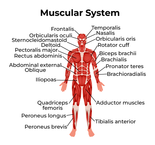
Some of the examples of skeletal muscles include:
- Biceps Brachii: The muscles in the upper arm that flex the elbow joint and supinate the forearm are called the biceps brachii.
- Quadriceps Femoris: This set of four muscles, which is situated in the front of the thigh, is responsible for flexing the hip joint and extending the knee joint.
- Gastrocnemius: Located in the leg’s calf area, the gastrocnemius is responsible for flexing the knee joint and plantar flexion of the foot.
- Rectus Abdominis: This muscle, which is located in the anterior abdominal wall, is responsible for compressing the internal organs of the abdomen and flexing the trunk.
- Trapezius: The trapezius, which is found in the neck and upper back, controls the scapula’s rotation, retraction, elevation, and depression.
Skeletal Muscles Properties:
The following are the properties of skeletal muscles:
- Voluntary Control: Skeletal muscles can be activated and regulated based on an individual’s purposes and needs.
- Striated Appearance: Under a microscope, the structured arrangement of actin and myosin filaments within muscle fibers gives skeletal muscle tissue a striped or striated appearance.
- Attachment to Bones: Tendons attach skeletal muscles to bones, allowing them to push against the skeleton and cause movement at joints.
- Excitability: Skeletal muscles can respond to stimuli from the nervous system or other muscle fibers by generating electrical impulses known as action potentials.
- Contractility: Skeletal muscles have the ability to shorten forcibly when stimulated, enabling them to produce movement by pulling on bones and other structures.
- Elasticity: After contraction, skeletal muscles can return to their original length due to their elastic properties, allowing for controlled movement and preventing overextension.
- Extensibility: Skeletal muscles can lengthen passively beyond their resting length when an external force is applied.
- Multinucleated Fibers: The skeleton mucles are multiplenucleated. This means it has multiple nuclei in the muscular fibers of skeletal muscle tissue promote the production and repair of proteins.
Skeletal Muscle Types:
Skeletal muscles can be divided into two types:
- Red Muscles: The red pigment known as myoglobin, which is present in large quantities in the human body, is responsible for the appearance of red muscles. These muscles have a greater number of mitochondria and a smaller diameter. The oxygen that the mitochondria require to synthesize ATP is stored in the myoglobin. There are a lot of blood capillaries in red muscles.
- White Muscles: The white muscles have a larger diameter and contain less myoglobin than the red muscles. Also, there are fewer mitochondria in them.